


Joint, Ganglion, and Bursal Aspiration in Warsaw
A joint, ganglion, or bursa aspiration allows for the removal of accumulated fluid, reduces the sensation of pressure, and collects material for further diagnostic testing. At CareLife Clinic in Warsaw, the procedure is preceded by an orthopedic consultation, during which the doctor assesses the nature of the condition and the indications for the procedure. Depending on the location, the aspiration may be performed under ultrasound guidance. We help, among others, patients with knee effusion, painful ganglions of the wrist, hand, or foot, and bursitis.
+340 Satisfied Patients
+12 Specialists on the team
+2,400 Procedures per year
+15 Years of experience
Joint, Ganglion, and Bursal Aspirationin Warsaw
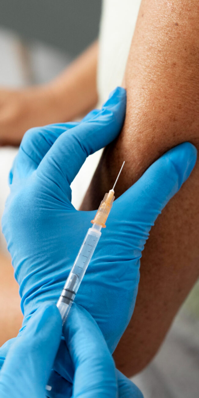
A puncture is a minimally invasive procedure that involves inserting a sterile needle into a joint, bursa, or hyaline cyst. It allows for the drainage of accumulated fluid, reduces tension and pain, and, in certain situations, enables the collection of material for laboratory testing. After draining the lesion, the doctor may decide to administer an appropriately selected medication.
At CareLife Clinic in Warsaw, we perform punctures for conditions such as knee joint effusion, bursitis, and symptomatic ganglions of the wrist, hand, or foot. Each procedure is preceded by a consultation and medical evaluation. Depending on the location of the lesion and the medical indications, the aspiration may be performed under ultrasound guidance.
What is an orthopedic puncture?
at CARELIFE CLINIC?
An orthopedic puncture, also known as aspiration, involves puncturing a specific structure and draining the fluid inside it. In the case of a joint, this may be excess synovial fluid, blood, or inflammatory fluid. From a ganglion, thick, gelatinous contents are removed, while from a bursa, fluid accumulating as a result of inflammation, injury, or chronic compression is removed.
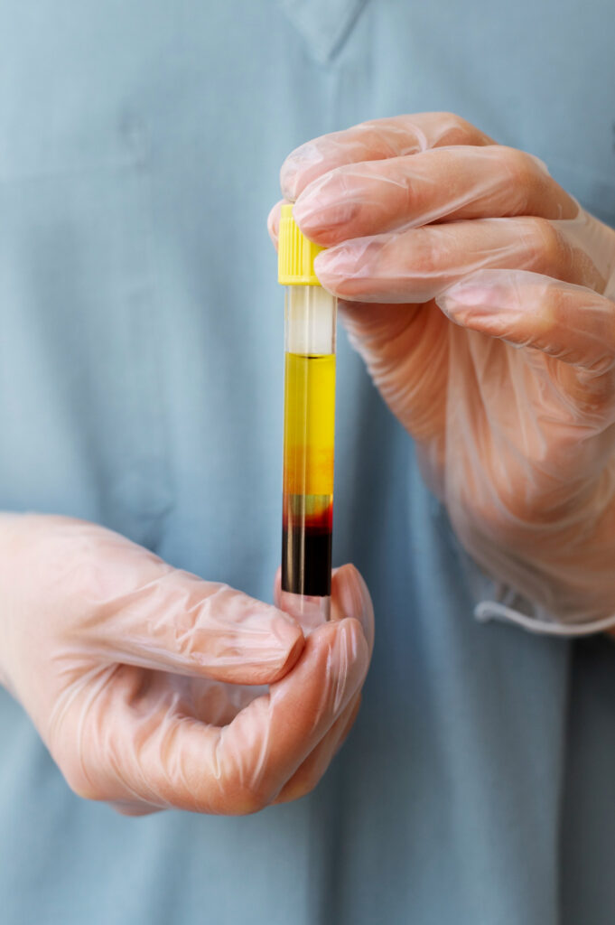
The procedure can be therapeutic or diagnostic. Drainage reduces tissue distension, swelling, and restricted mobility, but it does not always eliminate the underlying cause of the problem. The extracted material can be sent for analysis, for example, to check for infection, the presence of sodium urate or calcium pyrophosphate crystals, and the nature of the inflammatory process.
Specialists who perform orthopedic punctures at Carelife Clinic


The puncture is performed by a physician who specializes in the diagnosis and treatment of musculoskeletal disorders. Before the procedure, the specialist assesses whether the swelling is caused by joint effusion, a ganglion cyst, bursitis, or another condition requiring a different course of treatment. If necessary, the specialist uses an ultrasound examination to assess the location of the fluid and safely plan the puncture.
Experience with ultrasound-guided procedures is particularly important for small, deep-seated lesions and those located near blood vessels or nerves. After the aspiration, the physician determines the next course of action, which may include observation, follow-up, medication, rehabilitation, or referral for surgical treatment.
We’re open 7 days a week!
When is a joint aspiration performed?
A joint aspiration is considered when there is significant effusion, swelling, pain, and limited joint mobility. It may be necessary following an injury, in the course of osteoarthritis, gout, rheumatic diseases, or other inflammatory conditions. It is also performed when a doctor needs to determine why fluid has accumulated in the joint.
A joint that has suddenly become swollen, very painful, red, and noticeably warmer requires urgent evaluation, especially if the symptoms are accompanied by a fever or general malaise. In such a situation, a joint infection must be ruled out. A joint aspiration may then be part of the diagnostic process, but further management depends on the test results and the patient’s condition.
How should you prepare for a joint aspiration?
It’s a good idea to bring the results of previous tests, hospital discharge summaries, and reports from ultrasound, X-ray, or MRI scans to your appointment. You should prepare a list of medications and supplements you are taking, with particular attention to anticoagulants and antiplatelet agents. Do not stop taking them on your own—the decision to adjust the dosage, if necessary, is made by your doctor.
Before the procedure, you must inform the specialist of any fever, ongoing infection, skin lesions at the planned injection site, diabetes, bleeding disorders, or allergies to medications or disinfectants. The skin should be clean and free of creams and ointments. Fasting is usually not required, unless the doctor gives other instructions.
Orthopedic Injection in Warsaw - Price List
Maciej Kostewicz, M.D.
Joint/ganglion/bursal puncture – 250 zlotys
Are you thinking about choosing a service?
We can help you
We’re open 7 days a week!
Joint and Ganglion Aspiration in Warsaw’s Ochota District — a procedure at CareLife Clinic
CareLife Clinic is located at 3 Józefa Siemieńskiego Street in Warsaw’s Ochota district. At the clinic, patients can receive orthopedic and traumatology consultations, musculoskeletal diagnostics, and joint, ganglion, or bursa aspiration. This allows for the diagnosis of the problem, patient evaluation, and planning of further treatment all under one roof.
Depending on the indications, the aspiration may be performed under ultrasound guidance. After the procedure, the patient receives recommendations regarding dressing care, activity, and monitoring of the treated area. If drainage alone does not resolve the problem, care may be supplemented with further diagnostics, orthopedic treatment, or individually tailored rehabilitation.
Knee Aspiration and Joint Fluid Drainage
The knee is one of the joints most prone to fluid buildup. Effusion may occur following a sprain, contusion, meniscus or ligament injury, overuse, exacerbation of degenerative changes, or as a result of an inflammatory condition. The patient may experience a sensation of fullness, stiffness, pain when bending the knee, and difficulty fully straightening the leg.
During a knee aspiration, the doctor inserts a needle into the joint cavity and draws the fluid into a syringe. Removing a large amount of effusion can reduce tension in the joint capsule and make it easier to move the knee. However, drainage alone does not replace treatment of the underlying cause. Recurrent fluid buildup may require an ultrasound, X-ray, or MRI, as well as further orthopedic treatment or rehabilitation.
Ganglion Cyst Aspiration — Treatment of a Ganglion Cyst
A ganglion is a benign cyst filled with thick, gelatinous fluid. It usually forms near a joint or tendon sheath and may be connected to them by a narrow canal. Not every ganglion cyst requires treatment. Observation is often sufficient if the cyst is painless, does not grow, and does not limit limb function.
A puncture is considered when the lump causes pain, pressure, limited mobility, weakened grip, tingling, or significant discomfort. During the procedure, the doctor drains the contents of the cyst. It is important to note that the cyst’s walls and its connection to the joint may remain, so the ganglion may refill with fluid. Aspiration is a less invasive method than surgery, but it carries a higher risk of recurrence.
Bursal Aspiration
Bursae are small, fluid-filled structures that reduce friction between a bone and a tendon, muscle, or the skin. Due to injury, prolonged pressure, intense activity, or an inflammatory condition, excess fluid may accumulate in a bursa. Common locations include the elbow, kneecap, hip, and shoulder.
A bursal puncture allows for the drainage of fluid and, if necessary, the collection of a sample for testing. This is particularly important when the doctor suspects an infection or gout. Once an infection has been ruled out and if medically indicated, the use of an anti-inflammatory medication may be considered. In the case of an infected bursa, a different course of treatment is necessary, which may include antibiotic therapy and further medical follow-up.
Ganglion Cysts of the Wrist, Hand, and Foot
Ganglions most commonly appear on the dorsal or palmar side of the wrist, near the fingers, on the palm, and in the foot and ankle. They may vary in size, periodically disappear, and then enlarge again. Symptoms often worsen during manual labor, exercise, supporting oneself on the hands, running, or wearing tight shoes.
Before the aspiration, the doctor assesses the location of the lesion and its relationship to tendons, nerves, and blood vessels. This is particularly important in the case of a ganglion on the palmar side of the wrist, where the radial artery may run nearby. Not every lump is a ganglion, so in cases where the findings are inconclusive, an ultrasound may be necessary, and sometimes further diagnostic testing may be required.
Joint effusion, cysts, and bursitis—how do they differ?
Joint effusion refers to an increased amount of fluid in the joint cavity. It can result from trauma, overuse, osteoarthritis, gout, rheumatic disease, or infection. It most commonly causes diffuse swelling, a sensation of fullness, and limited mobility of the entire joint.
A ganglion is a localized cyst containing a gelatinous substance, usually visible as a localized lump. A bursa, on the other hand, is located outside the joint cavity and facilitates the gliding of tissues. Its inflammation causes localized swelling and tenderness, such as on the back of the elbow or in front of the kneecap. An accurate diagnosis determines the puncture site, the purpose of the aspiration, and further treatment.
Ultrasound-guided aspiration
Ultrasound allows the doctor to observe the needle and its position in real time. It allows for the precise identification of the site of fluid accumulation, assessment of the size and structure of the lesion, and planning of a safe puncture path. It also makes it easier to avoid nearby tendons, blood vessels, and nerves.
Ultrasound guidance is particularly useful for small, deep-seated, or multilocular lesions, as well as in areas with complex anatomy. It increases the precision of the puncture but does not guarantee that the effusion or ganglion will not recur. The risk of recurrence depends primarily on the cause of the fluid accumulation and the nature of the lesion being treated.
Orthopedic Consultation and Qualification for Aspiration
Before the procedure, the doctor takes a medical history, examines the painful area, and reviews the patient’s medical records. The doctor assesses, among other things, the duration of symptoms, previous injuries, the rate at which the swelling is increasing, limited movement, and the presence of fever, redness, or neurological symptoms.
The evaluation also takes into account medications being taken, chronic conditions, coagulation disorders, allergies, and previous punctures or surgeries. If the clinical picture is unclear, the doctor may perform or order an ultrasound, X-ray, or MRI. A puncture is not performed automatically for every swelling—it must first be determined whether the procedure will be safe and truly necessary.
Ganglion Cyst Aspiration vs. Surgical Removal
A puncture is a minimally invasive procedure; it does not require an incision in the skin and usually allows for a quick return to daily activities. Its limitation is that it leaves the cyst wall intact, along with any possible canal connecting it to the joint or tendon sheath. For this reason, the ganglion may refill with fluid.
Surgery involves excising the cyst along with its stalk, but it is a more extensive procedure, requires wound healing, and carries a risk of scarring, stiffness, and damage to nearby structures. Surgical treatment is more often considered for recurrent or multichambered lesions that cause nerve compression, functional impairment, or persistent symptoms despite conservative treatment.
Returning to Normal Activities After a Puncture
After an uncomplicated puncture, many people can resume light activities on the same day. Office work is usually possible immediately or the next day, provided that pain and swelling do not interfere with performing duties. After a lower-limb puncture, your doctor may temporarily recommend limiting long walks.
A return to exercise should be gradual. In most cases, light activity can be resumed after 24–48 hours, while more strenuous exercise should be postponed for a few days and made contingent on the body’s response. If the effusion was caused by overuse or a biomechanical disorder, physical therapy focused on joint mobility, strength, and control may be helpful.
Contraindications to puncture
The most important contraindication is a skin infection at the planned puncture site, as the needle could introduce microorganisms into deeper tissues. A scheduled procedure is also not performed during an acute infection accompanied by fever. However, if an infection of the joint itself or the bursa is suspected, a puncture may be an urgent part of diagnosis and treatment—in which case it requires appropriate medical management.
Coagulation disorders, a significantly reduced platelet count, and the use of anticoagulants or antiplatelet medications require special consideration. These medications do not always preclude a puncture, but the doctor must assess the risk of bleeding on a case-by-case basis. Other contraindications may include a lack of safe access to the lesion or a lack of confirmation that the observed lump is indeed a ganglion.
Possible Side Effects and Warning Signs
After the aspiration, you may experience short-term pain, tenderness, slight swelling, bruising, or a trace of blood on the dressing. Less commonly, more severe bleeding, infection, a reaction to the administered medication, or damage to a nearby nerve, blood vessel, or tendon may occur. The use of sterile technique and ultrasound guidance when appropriate helps reduce the risk but does not eliminate it entirely.
The following symptoms require immediate medical attention: pain that worsens rather than subsides, fever, chills, rapidly enlarging swelling, intense redness and warmth of the skin, purulent discharge, numbness in the limb, muscle weakness, or a marked deterioration in mobility. Sudden paleness, bruising, or cooling of the limb requires immediate medical evaluation.
Frequently Asked Questions About Joint, Ganglion, and Bursal Aspiration
Below, we answer patients’ most common questions regarding preparation for the puncture, the procedure itself, analysis of the fluid sample, return to normal activities, and the risk of recurrence. Final recommendations always depend on the site of the procedure, the diagnosis, and the patient’s overall health.
How long does the aspiration take?
The procedure itself usually takes a dozen or so minutes. More time should be set aside for the consultation, examination, preparation of the sterile field, and any necessary ultrasound.
What happens during a joint, ganglion, or bursa aspiration?
The patient assumes a position that allows for easy access to the treatment area. The doctor disinfects the skin, prepares a sterile field, and—depending on the location and type of procedure—may administer local anesthesia. The doctor then inserts a needle into the joint, bursa, or cyst, drains the fluid, and assesses its appearance.
If there are diagnostic indications, the collected material is sent to the laboratory. After ruling out any contraindications, the doctor may administer an appropriate medication, but this is not a mandatory part of every aspiration. After the needle is removed, a dressing is applied, sometimes a compression bandage. The procedure itself usually takes about a dozen or so minutes, although the consultation, ultrasound, and preparation extend the total duration of the visit.
Does the aspiration hurt?
The patient may feel a prick when the needle is inserted and a brief sensation of pressure or tightness while the fluid is being drained. The severity of these sensations depends on the puncture site, the size of the lesion, and the degree of inflammation. Local anesthesia is used if necessary.
After the procedure, the area may remain tender for several hours or days. A slight bruise or temporary discomfort when moving the limb usually does not indicate a complication. However, the pain should gradually subside. A marked increase in pain, especially accompanied by fever, redness, or worsening swelling, requires consultation with a doctor.
What can be administered after the aspiration?
After draining the fluid, the doctor may conclude the procedure without administering any additional medication. In certain non-infectious inflammatory conditions, the administration of a glucocorticosteroid is considered, sometimes in combination with an anesthetic. The decision depends on the diagnosis, the condition of the tissues, the patient’s medical history, and prior treatment.
Hyaluronic acid or platelet-rich plasma (PRP) are separate forms of therapy and are not routinely administered after every aspiration. They require specific indications and patient eligibility criteria. If an infection is suspected, administering a steroid preparation into the joint or bursa is contraindicated—the first step is to diagnose the aspirated fluid and treat the cause of the infection.
When is a puncture not enough?
Draining the fluid may reduce symptoms, but it will not repair a damaged meniscus, ligament, cartilage, or advanced degenerative changes. If the effusion returns quickly, the cause must be investigated. Additional tests, medication, physical therapy, or surgery may be necessary.
Aspiration may also be insufficient in cases of multicameral or recurrent ganglion cysts, joint or bursal infections, or when the lesion is compressing a nerve or blood vessel. In such situations, the doctor determines the next course of treatment. Subsequent punctures without diagnosing the cause of the recurrences should not replace a comprehensive orthopedic evaluation.
Can the fluid or ganglion come back?
Yes. A joint aspiration removes the accumulated fluid, but it does not always eliminate the underlying cause. Joint effusion may recur if inflammation, overuse, intra-articular damage, or an active systemic disease persists. Similarly, a bursa may refill if it remains exposed to pressure or repetitive trauma.
Recurrence is also common after ganglion aspiration, as the cyst wall and its connection to the joint may remain. This does not mean that the procedure was performed incorrectly. If the lesion reappears, the doctor will assess whether further observation, a repeat aspiration, modification of activity, rehabilitation, or surgical treatment is indicated.